
Author Archives: Stephen Huffman

AED Pads for Adults: Types, Placement, and When to Replace Them

Adult AED pads are adhesive electrode pads placed on the bare chest of a person experiencing sudden cardiac arrest. They connect the patient to the AED, allowing it to analyze the heart rhythm and deliver a life-saving electrical shock. For adults and anyone over 8 years old or weighing more than 55 pounds, specific adult-size pads and placement positions are always required.

Key Takeaway

  • Adult AED pads are essential for delivering a life-saving shock during sudden cardiac arrest, and correct placement helps ensure effective defibrillation. Always follow the AED’s instructions and use pads compatible with the specific device. Check expiration dates regularly and replace pads after every emergency use or if the packaging is damaged. Understanding adult and pediatric pad differences, along with special placement considerations, helps you respond more confidently in an emergency. Regular AED inspections and hands-on CPR/AED training help keep you prepared when every second counts.

What Are Adult AED Pads?

AED electrode pads, sometimes called defibrillator pads or electrodes, are the critical interface between an automated external defibrillator and the patient's heart. Each pad is coated with a conductive adhesive gel that allows the device to both read the heart's electrical activity and deliver a controlled shock when a shockable rhythm, such as ventricular fibrillation, is detected.

Adult AED pads are designed to deliver a higher-energy shock appropriate for a fully developed cardiovascular system. The American Heart Association (AHA) guidelines specify that adult pads should be used on any person aged 8 years or older or weighing more than 55 pounds (25 kilograms), regardless of age. Every AED sold in the United States must be FDA-approved, which means the pad adhesive, energy delivery parameters, and labeling all meet established safety standards.

Because AED pads are single-use consumables, they must be replaced after every emergency deployment (even if no shock was delivered) and before their printed expiration date. This is a detail that surprises many new AED owners: the pads expire on schedule regardless of whether the device has ever been used.

Types of Adult AED Pads:The Guide No E-Commerce Page Gives You

Most guides skip this section entirely. Understanding the different types of adult AED pads before an emergency occurs is one of the most underrated elements of AED preparedness. It is exactly the kind of knowledge that differentiates a trained responder from an untrained bystander.

Standard Electrode Pads

Standard electrode pads are the most widely used type. They consist of two separate adhesive pads connected to the AED unit via cables. You manually peel each pad from its backing and press it firmly onto the appropriate position on the patient's bare chest. Standard pads are the default configuration for most AED brands, including ZOLL STAT-PADZ, Defibtech Lifeline pads, and Cardiac Science PowerHeart pads. These pads are available for both adults and children and are the most straightforward to stock and replace.

Pre-Connected Pads

Pre-connected pads come already attached to the AED unit with cables, eliminating the step of connecting the pad cable during an emergency. Philips HeartStart models, including the FRx, use a pre-connected pad cartridge system. When the device is opened and turned on, the pads are immediately ready to apply, removing one step from an already high-stress situation. This design is particularly valuable in public-access settings where untrained bystanders may need to act quickly.

Universal / Uni-Pads

Universal pads, sometimes called uni-pads, are designed to work on both adults and children by automatically adjusting the energy level delivered based on pad-detected signals or a manual switch. ZOLL AED 3 and certain Philips FRx models support universal pad configurations. Uni-pads eliminate the need to carry two separate pad sets and reduce the risk of grabbing the wrong pads in an emergency. For organizations that serve both adults and children (schools, community centers, fitness facilities) this is an important capability to know before purchasing.

Training Pads

Training pads are semi-adhesive, reusable pads designed exclusively for use with AED trainer units in practice scenarios. They simulate the feel and application of real pads without consuming rescue supplies. SimpleCPR's Adult CPR/AED course [LINK → https://www.simplecpr.com/adult-cpr-aed] teaches proper pad application and placement as part of AED certification. One reason hands-on practice with training pads builds the muscle memory that matters in a real emergency. Training pads should never be used with a live AED on a patient.

CPR Feedback Pads

Some advanced AED pad models include built-in CPR feedback sensors. AED.com notes that certain electrode pads are designed to provide real-time CPR guidance, monitoring compression depth and rate while defibrillation is being prepared or performed. ZOLL's CPR-D-Padz are the most widely recognized example of this design. For healthcare providers and high-performance CPR programs, feedback-enabled pads provide continuous quality monitoring alongside defibrillation capability.

How to Place AED Pads on an Adult

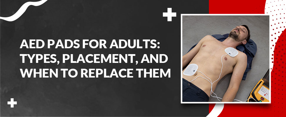
Correct pad placement helps ensure the electrical current travels through the heart muscle via the most direct pathway, maximizing the chance of restoring a normal rhythm. Every AED sold with adult electrode pads includes placement diagrams printed directly on the pads: follow those diagrams as your first reference. The following applies to any adult victim aged 8 or older.

IMPORTANT: Do not delay defibrillation to resolve placement uncertainty. Survival rates drop 7–10% for every minute without a shock. If the pads are approximately in position, proceed. The AED's own prompts will guide you.

Anterolateral Placement: The Standard Adult Position

Anterolateral placement is the default method recommended by the AHA for all adult victims. "Anterior" means front; "lateral" means side. Follow these steps:

  • Step 1: Expose the chest. Remove or cut away clothing. AED pads will not function through fabric.

  • Step 2: Dry the chest. If the person is wet, quickly wipe the chest with a cloth or towel. Moisture reduces pad adhesion and can interfere with shock delivery.

  • Step 3: Check for obstructions. Look for excessive chest hair (shave quickly with the razor included in most AED kits), medication patches (remove them), or an implanted pacemaker/ICD (visible as a small bump under the skin: see special situations below).

  • Step 4: Place pad 1 (right chest). Position the first pad on the upper right side of the chest, just below the right collarbone (clavicle), to the right of the sternum (breastbone).

  • Step 5: Place pad 2 (left side). Position the second pad on the lower left side of the chest, below the armpit along the mid-axillary line, just below the left nipple.

  • Step 6: Connect and follow prompts. Plug in the cable connector if not pre-connected. Turn on the AED and follow its voice and visual prompts.

  • Step 7: Clear and analyze. Ensure no one is touching the patient while the AED analyzes the heart rhythm. If a shock is advised, ensure all bystanders are clear before pressing the shock button.

Anterior-Posterior Placement

Anterior-posterior (AP) placement positions one pad on the front of the chest and the other on the back. This method is most commonly used in clinical settings. It is also recommended when placing pads near an implanted pacemaker or ICD, or in certain obesity scenarios where AP placement provides better pad contact.

For AP placement on adults: position the anterior pad on the center of the left chest, over the cardiac apex. Position the posterior pad at the center of the back, between the shoulder blades, directly behind the anterior pad. Ensure both pads lie flat against bare skin and do not overlap or touch.

PLACEMENT COMPARISON TABLE:

Feature

Anterolateral

Anterior-Posterior

When to Use

Pad positions

Right chest + lower left side

Front chest + center back

—

AHA default?

✓ Yes, standard for adults

No, alternative method

—

Best for adults?

Yes, all adult victims

Yes, specific situations

See notes below

Use when the pacemaker is near the right shoulder?

Move the pad at least 1 inch away from the device

✓ Preferred alternative

Pacemaker / ICD present

Clinical use?

Yes

✓ Often preferred in clinical settings

Manual defibrillators, hospital

Pad overlap risk?

Low, pads are on different sides.

Low if positioned correctly

—

Special Situations for Adult AED Pad Placement

Standard anterolateral placement applies to the vast majority of adult victims. However, a handful of common situations require adjustments. Knowing these before an emergency removes hesitation.

Pacemaker or ICD

A pacemaker or implantable cardioverter-defibrillator (ICD) is visible as a small lump under the skin, typically in the upper chest area. Do not place an AED pad directly over the device as the metal housing can divert the shock away from the heart. Position the pad at least 1 inch (2.5 cm) away from the device. If the pacemaker is located where the right pad would normally go, use anterior-posterior placement instead.

Medication patches

Transdermal patches (nitroglycerin, nicotine, hormones) must be removed before applying pads. Leaving a patch under an electrode can cause skin burns and may interfere with current delivery. Wear gloves when removing a patch to avoid absorbing the medication yourself.

Wet chest

Moisture creates resistance and reduces pad adhesion. Quickly dry the chest with whatever is available (a shirt, towel, or jacket) before applying pads. Do not delay this step by more than a few seconds.

Excessive chest hair

Dense chest hair prevents pads from adhering to the skin. Most AED response kits include a disposable razor. Shave the specific area where each pad will be placed. If a razor is unavailable, press the pads down firmly and then peel them off quickly (the adhesive will pull some hair away), and then apply the second pair of replacement pads. This is why keeping a spare set of pads is always recommended.

Pregnant women

AED pad placement for pregnant women is identical to standard adult anterolateral placement. The AHA confirms that defibrillation does not harm the fetus and must not be delayed. Restoring the mother's heart rhythm is the highest priority for both lives.

Large breasts

The lateral pad should be positioned beneath the breast tissue, not on top of it. Direct skin contact is required for effective defibrillation.

Adult vs. Pediatric AED Pads: Key Differences

One of the most important questions in AED readiness is knowing when to use adult pads and when to use pediatric pads, and what to do when you have only one type available.

Factor

Adult AED Pads

Pediatric AED Pads

Designed for

Adults and children ≥ 8 years or ≥ 55 lbs

Children < 8 years or < 55 lbs (25 kg)

Energy level

Higher joule output, appropriate for the adult heart

Attenuated / reduced joule output (safe for pediatric heart)

Pad size

Larger

Smaller, designed for a smaller chest wall

Default placement

Anterolateral (right chest + lower left side)

Anterior-posterior (front chest + back)

If only adult pads are available for a child?

Use adult pads (a shock with adult pads is better than no shock). Position front-to-back to prevent overlap.

N/A

Can pediatric pads be used on adults?

No. AHA guidelines advise against it. The pediatric energy dose is too low for an adult heart. Do not substitute.

N/A

The critical takeaway: if no pediatric pads are available, use adult pads on a child rather than doing nothing. But never reverse this and use child pads on an adult victim. The energy delivered will almost certainly be insufficient to convert the arrhythmia.

AED Pad Expiration and Replacement: A Schedule That Saves Lives

AED pads expire because the conductive adhesive gel they rely on degrades over time. Once the gel dries out or loses adhesion, the pads may not stick properly to the patient's chest, creating resistance that reduces or blocks the shock's effectiveness. An AED with expired pads may still power on and appear functional, but it cannot be relied upon in a rescue.

Expired pads are one of the leading causes of AED failure when a device is actually needed. Routine maintenance prevents this entirely.

AED Brand / Model

Adult Pad Shelf Life

Battery Life

Notes

Philips HeartStart OnSite / HS1

~2 years

~4 years

SMART Pads Cartridge (M5071A) with expiry printed on package

ZOLL AED Plus

CPR-D-Padz: ~5 years; STAT-PADZ II: ~2 years

~5 years

Two pad options available, so confirm which your unit uses

HeartSine Samaritan PAD

~4 years (Pad-Pak combined)

~4 years (Pad-Pak combined)

Battery and pads in one cartridge, so one expiration to track

Defibtech Lifeline

~2 years

5–7 years (model dependent)

Always replace pads immediately after any emergency use

Cardiac Science Powerheart

~2 years

Per model spec

Check packaging (date printed on cartridge label)

REPLACE PADS IMMEDIATELY if any of the following apply:

  • The printed expiration date on the package has passed.

  • The pads were applied to a patient during an emergency (even if no shock was delivered).

  • The pad package has been opened, torn, punctured, or shows signs of moisture damage.

  • The AED's status indicator is showing a warning or fault rather than a ready signal.

Best practice for organizations: conduct a monthly visual inspection of each AED. Confirm the status indicator is green, check the expiration date on the pad package, and order replacements at least 60 days before expiration to avoid gaps in coverage.

AED Pads and CPR Certification: What Every Trainee Needs to Know

Understanding AED pads on paper is valuable. Applying them under pressure (when someone has collapsed, bystanders are panicking, and every second counts) is a different skill entirely. This is why hands-on AED training matters.

In SimpleCPR's Adult CPR/AED certification course, students learn AED pad application as an integrated component of the full chain-of-survival response: recognizing cardiac arrest, calling 911, starting CPR compressions, and deploying the AED without interrupting chest compressions for longer than necessary. The AED prompts guide the rescuer through analysis and shock delivery, but trainees who have practiced the physical steps of pad application are measurably faster and more confident.

Key skills the certification covers related to AED pads:

  • Identifying which pads to use (adult vs. pediatric) for the victim in front of you.

  • Applying pads to bare skin while another responder maintains chest compressions.

  • Recognizing and responding to common complications: wet chest, hairy chest, visible pacemaker.

  • Coordinating the "all clear" call before shock delivery to protect bystanders.

  • Resuming CPR immediately after shock delivery without waiting for the AED's next analysis cycle to complete.

National data consistently show that bystander AED use before emergency services arrive significantly increases cardiac arrest survival rates. CPR and AED certification empowers anyone (regardless of medical background) to act with confidence in those first critical minutes. SimpleCPR's courses are completed in 60 minutes or less, are based on the latest AHA guidelines, and can be accessed 24 hours a day.
Stroke vs. Heart Attack: What Every Bystander Needs to Know

A stroke and a heart attack are both medical emergencies caused by disrupted blood supply, but they affect different organs. A heart attack cuts off blood to the heart muscle; a stroke cuts off blood to the brain. Their symptoms, treatments, and emergency responses differ. For trained bystanders, knowing which condition you are dealing with determines the actions you take next.

Key Takeaway

  • Understanding the difference between a stroke and a heart attack helps bystanders recognize symptoms and respond appropriately. Both conditions require immediate emergency medical attention, as every minute can affect survival and recovery. Recognizing warning signs, noting when symptoms begin, and following emergency dispatcher instructions can save lives. Knowing when to perform CPR and why aspirin should be avoided in suspected strokes is equally important./li>

What Is a Heart Attack?

A heart attack (medically known as a myocardial infarction) happens if a coronary artery becomes blocked, most commonly by a blood clot that forms over a ruptured atherosclerotic plaque. The heart muscle supplied by that artery is starved of oxygen. Without restored blood flow, heart cells begin dying permanently within minutes.

One critical point every bystander should understand: a heart attack does not mean the heart has stopped beating. The heart continues to function, but it is being damaged by an interruption in its own blood supply. This is fundamentally different from cardiac arrest, which is addressed in a later section of this article.

Heart Attack Symptoms

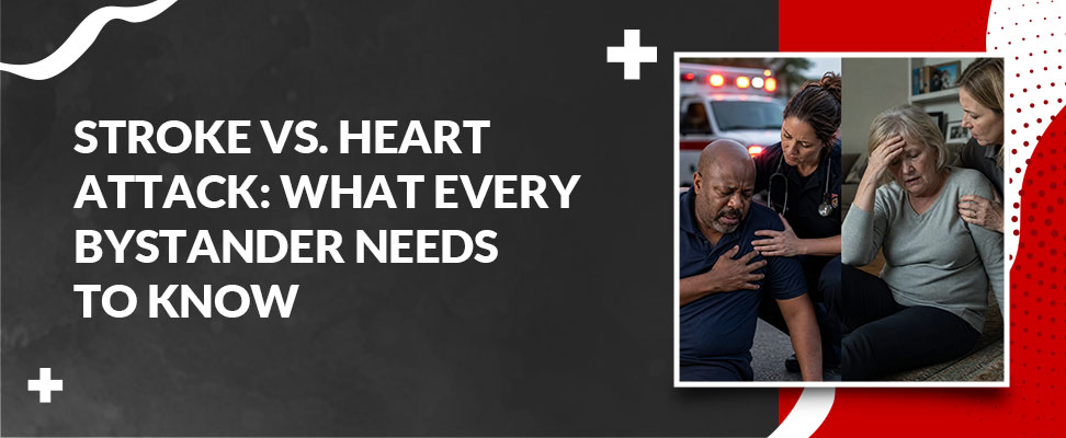
Heart attacks do not always present as dramatic, movie-style chest clutching. Common symptoms include:

  • Chest discomfort: pressure, squeezing, tightness, or pain often described as "an elephant on my chest," which often pulses through the left arm, jaw, neck, or back.

  • Shortness of breath sometimes appears before chest discomfort begins.

  • Nausea, lightheadedness, or a sudden cold sweat.

  • Unusual or extreme fatigue: particularly in women, which can begin hours or days before the event.

  • Sense of impending doom or unexplained anxiety.

Some heart attacks are "silent," with no chest pain at all. People with diabetes are especially prone to atypical or minimal symptoms due to autonomic neuropathy affecting pain perception.

What Is a Stroke?

A stroke happens if blood flow to a region of the brain is interrupted. Brain cells require a constant oxygen supply and begin dying within minutes of that supply being cut off. Unlike the heart muscle, which can partially recover function, brain tissue that dies does not regenerate, making the speed of treatment even more critical.

Two Types of Stroke: Ischemic and Hemorrhagic

Ischemic strokes are usually 87% of all strokes. It is caused by a blood clot blocking an artery supplying the brain. The clot may form locally due to atherosclerosis or travel from elsewhere in the body, particularly from the heart in people with atrial fibrillation (AFib).

Hemorrhagic stroke accounts for the remaining 13% and happens due to a ruptured blood vessel bleeding into or around the brain. The resulting pressure damages the surrounding brain tissue. High blood pressure is the most common underlying trigger.

A transient ischemic attack (TIA), sometimes called a mini-stroke, has stroke-like symptoms that resolve within 24 hours. It is not minor: up to 15% of people who experience a TIA have a full stroke within three months. TIA requires emergency evaluation, not a "wait and see" approach.

Stroke Symptoms: Use BE-FAST

The BE-FAST acronym gives bystanders a reliable framework for stroke recognition:

  • B (Balance): Sudden loss of coordination or ability to walk normally.

  • E (Eyes): Sudden vision loss, blurring, or double vision in one or both eyes.

  • F (Face): Drooping on one side. Ask the person to smile- does one side fall?

  • A (Arms): Weakness in one arm. Ask them to lift up both arms- does one drift down?

  • S (Speech): Slurred, confused, absent, or strange speech. Ask them to repeat a phrase.

  • T (Time): Call 911 immediately if any signs are present. Note the exact time symptoms began.

That last point is critical: the time of symptom onset determines whether clot-dissolving medication (tPA) can be safely administered. The treatment window is 4.5 hours from the first symptom for most ischemic strokes.

Stroke vs. Heart Attack: Key Differences at a Glance

Organ Affected

Heart

Brain

Primary mechanism

Blocked coronary artery

Blocked or ruptured brain artery

Hallmark symptom

Chest pressure / radiating pain

Facial droop, arm weakness, speech difficulty

Is the heart still beating?

Yes, but the muscle is being damaged

Yes, stroke does not stop the heart

CPR needed at onset?

NO, only if cardiac arrest develops

NO, Stroke does not cause cardiac arrest

AED appropriate?

Only if cardiac arrest (VF/pulseless VT) develops

NO, AEDs have no role in stroke

Give aspirin?

YES, chew 325mg if conscious, no allergy

NO, dangerous if hemorrhagic stroke

Clot-dissolving drug?

Aspirin, anticoagulants, PCI procedure

tPA within 4.5 hrs for ischemic only; NOT for hemorrhagic

Treatment motto

"Time is muscle"

"Time is brain"

Treatment time window

Restore blood flow as fast as possible

4.5 hrs for tPA; thrombectomy up to 24 hrs in some cases

Shared Risk Factors

Despite affecting different organs, strokes and heart attacks share a remarkably similar set of modifiable risk factors. Reducing these risks simultaneously lowers the probability of both conditions.

  • High blood pressure (hypertension) is a crucial modifiable risk factor for both conditions. Sustained elevated pressure accelerates arterial wall damage and dramatically increases the risk of clot formation.

  • Atherosclerosis: Plaque buildup inside arterial walls narrows vessels and creates vulnerable rupture sites in both coronary and cerebral arteries.

  • Atrial fibrillation (AFib): This irregular heart rhythm causes blood to pool in the heart's chambers, forming clots that can travel to coronary arteries (causing a heart attack) or to the brain (triggering a cardioembolic stroke).

  • Diabetes: Chronically elevated blood sugar levels damage blood vessel walls and accelerate atherosclerosis throughout the cardiovascular system.

  • Smoking, high LDL cholesterol, obesity, and physical inactivity all accelerate arterial damage, promote inflammation, and increase the risk of clot formation.

Addressing these factors, particularly blood pressure control and smoking cessation, is among the most impactful steps for preventing both conditions at once.

What Should You Actually Do as a Bystander?

This is the question most medical articles do not answer, and it is the most important question for anyone with first aid or CPR training: does your response depend on whether you think it is a stroke or a heart attack?

The answer is yes, and two of the most critical differences involve aspirin and CPR. Giving aspirin incorrectly, or withholding CPR at the wrong moment, can worsen outcomes. Here is the bystander decision framework.

If You Suspect a Heart Attack

  • Call 911 immediately. Do not drive the person to the hospital- paramedics can begin life-saving intervention during transport.

  • Help the person into a comfortable position. Sitting upright with knees bent often eases breathing. Loosen any tight clothing around the chest or neck.

  • Offer aspirin if appropriate. If the person is conscious, has no known aspirin allergy, and can swallow safely, have them chew (not swallow whole) one regular 325mg aspirin. Chewing speeds absorption. This may help slow clot formation in the coronary artery.

  • Do NOT start CPR unless the person loses consciousness and stops breathing normally. A heart attack is not the same as cardiac arrest- the heart is still beating. Beginning chest compressions on a conscious patient who still has a pulse is inappropriate.

  • Be prepared to use an AED if one is nearby, but only if the person becomes unresponsive and pulseless. An AED is not used on a conscious patient.

  • Record the exact time symptoms began and report this clearly when 911 connects.

If You Suspect a Stroke

A stroke requires a different set of actions, several of which are the direct opposite of what you would do for a heart attack:

  • Call 911 immediately. The very first thing you say to the dispatcher should be the time symptoms started- this determines which treatments are available at the hospital.

  • Do not give anything by mouth. Stroke frequently impairs the swallowing reflex, making food, water, or aspirin aspirational risks. More critically, never give aspirin to a suspected stroke patient. If the stroke is hemorrhagic, aspirin can significantly worsen the bleeding.

  • Use the recovery position if the person is unconscious but breathing. Place them on their side with their head slightly tilted back to protect the airway from saliva or vomit.

  • Do NOT perform CPR unless the person is unresponsive and not breathing normally. A stroke does not stop the heart from beating.

  • Do NOT deploy an AED. An AED corrects shockable heart arrhythmias. Stroke is a brain event, and it does not produce a shockable rhythm. An AED has no therapeutic role in stroke.

  • Keep the person calm and still. Minimize movement, particularly for hemorrhagic stroke.

If You Are Unsure (And Why That Is Okay)

In the field, it is not always possible to determine within the first minutes whether someone is having a stroke or a heart attack. If you are uncertain:

  • Call 911 first, without any delay.

  • Hold aspirin unless chest pressure is the primary symptom and there are no neurological signs (facial drooping, speech difficulty, limb weakness).

  • Start CPR only if the person is unresponsive and not breathing normally. This criterion applies regardless of which condition you suspect.

  • Describe all symptoms and their onset time accurately to the dispatcher and follow their instructions precisely.

Emergency dispatchers are trained to guide bystanders through exactly this uncertainty. Your role is to call early, report accurately, and follow dispatcher guidance.

Heart Attack vs. Cardiac Arrest: Not the Same Thing

One of the most dangerous misconceptions in emergency medicine is treating "heart attack" and "cardiac arrest" as interchangeable. They are distinct events with distinct responses.

What is happening

Coronary artery blocked; heart muscle losing blood supply

Heart stops beating effectively; no blood pumped

Is person conscious?

Usually yes, at least initially

No, sudden collapse and unresponsiveness

Is the person breathing?

Usually yes

No or only agonal gasping

Has the heart stopped?

No

Yes, cardiac output has ceased

Is CPR needed?

Only if cardiac arrest develops

Yes, begin immediately

Is an AED needed?

Only if cardiac arrest develops

Yes, deploy as fast as possible

Can one lead to the other?

Yes, heart attack can trigger ventricular fibrillation → cardiac arrest

–

A heart attack leads to cardiac arrest when the damaged heart muscle disrupts the organ's electrical system, producing ventricular fibrillation- a chaotic rhythm that generates no effective pumping. This escalation can occur without warning. It is the reason why calling 911 immediately at the first signs of a heart attack is so critical: the situation can deteriorate rapidly.

Can a Heart Attack Cause a Stroke?

Yes, though it occurs in a minority of cases. A heart attack degrades the heart muscle or triggers atrial fibrillation, both of which create conditions conducive to blood clot formation within the heart chambers. If a clot is dislodged and passes through the bloodstream into a cerebral artery, it can prevent blood flow to the brain, leading to a cardioembolic ischemic stroke. Research shows that stroke risk is meaningfully elevated in the first month following a heart attack, which is why anticoagulant therapy is often prescribed during cardiac recovery.

The reverse pathway is also documented. In brain regions that regulate cardiovascular autonomic function, such as the insular cortex, severe stroke can trigger a cascade of stress responses known as Stroke-Heart Syndrome. It potentially causes arrhythmias, myocardial injury, or sudden cardiac death. While both directional risks are real, they represent a minority of cases rather than the typical course.

Women's Symptoms: What May Present Differently

Both heart attack and stroke can present differently in women than in men, and these differences have historically contributed to delayed diagnosis and poorer outcomes.

Women Experiencing a Heart Attack May Have

  • Unusual or extreme fatigue, sometimes beginning days before the cardiac event itself.

  • Nausea, indigestion-like discomfort, or upper back pain rather than classic chest pressure.

  • Jaw pain, throat tightness, or shortness of breath without prominent chest symptoms.

  • A sudden, unexplained sense of anxiety or impending doom without a clear physical cause.

Women Experiencing a Stroke May Have

  • General weakness or malaise rather than clear one-sided paralysis.

  • Sudden, severe headache (especially prominent in hemorrhagic stroke).

  • Sudden confusion, agitation, or behavioral changes.

  • Hiccups or nausea are less commonly associated with stroke in public awareness materials.

Because these presentations differ from the classic descriptions most widely taught, women are statistically more likely to delay seeking emergency care. The rule for bystanders is consistent regardless of presentation: if something feels suddenly and inexplicably wrong, especially in someone with known cardiovascular risk factors, call 911 immediately and let emergency professionals evaluate.

What Is Pronator Drift? FAST Stroke Assessment Method Explained

Maybe a nurse mentioned it during a hospital visit. Maybe you read it in a textbook, or heard it on a medical show, and now you are wondering what pronator drift actually means. Here is the short version. Pronator drift is a clinical test doctors and nurses use to catch subtle arm weakness, often as an early clue to stroke.

But if you are the kind of person who wants to know what to actually do when someone in front of you might be having a stroke, the term you need is not pronator drift. It is FAST. We will cover what pronator drift means, what it can point to, and then get you to the test that matters most if you are the one standing there when it counts.

Key Takeaway

  • Pronator drift is a clinical neurological test used by healthcare professionals to detect subtle arm weakness that may indicate a stroke or other neurological condition. Bystanders should use the FAST test instead, checking for Face drooping, Arm weakness, Speech difficulty, and Time to call 911, to quickly recognize stroke symptoms and get emergency medical help without delay./li>

What Is Pronator Drift?

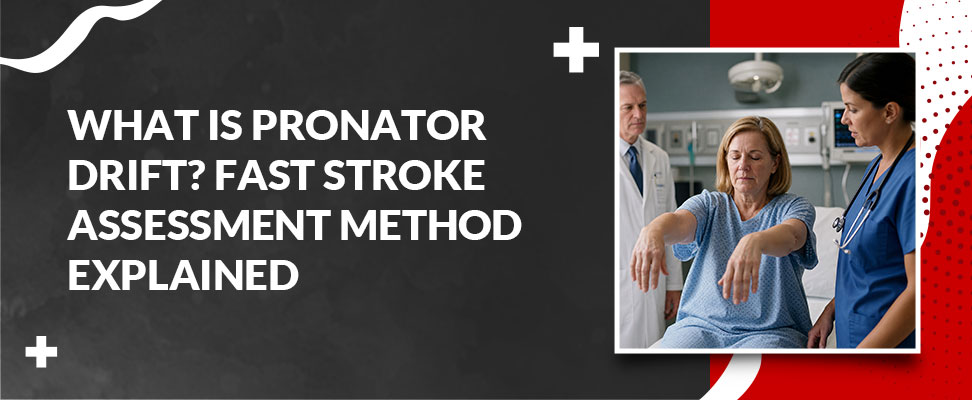
Pronator drift is a clinical neurological exam. The patient extends both arms straight out in front of them, parallel to the floor, with palms facing up, then closes their eyes and holds that position for 20 to 30 seconds while a clinician watches closely.

A positive result happens when one arm, or occasionally both, starts to drift downward while the palm rotates inward, or pronates. That inward turn is why the test carries the name it does. Even a small amount of drift or rotation counts as positive, which is exactly what makes this exam useful. It is built to catch weakness so mild that a standard strength test, where a clinician asks a patient to push or squeeze against resistance, might miss it entirely.

What Does a Positive Pronator Drift Mean?

A positive pronator drift points to a problem somewhere along the corticospinal tract, the pathway that carries movement signals from the brain down through the spinal cord to the muscles. When that pathway is disrupted, clinicians call it an upper motor neuron issue, and it tends to show up first as this kind of subtle, one-sided weakness.

The most common cause is stroke. Because of how the nervous system crosses over, a positive drift usually shows up on the side of the body opposite the affected side of the brain. A weak right arm, for example, often points to an issue on the left side of the brain.

Stroke is not the only explanation. Clinicians also use pronator drift to help evaluate multiple sclerosis, traumatic brain injury, brain tumors, and spinal cord lesions. On its own, a positive test is a sign, not a diagnosis. It tells a clinician where to look next, and it gets paired with the rest of the neurological exam, patient history, and often brain imaging before anyone concludes.

Who Performs This Test and Why It Is Not a Bystander Tool

Pronator drift is performed by trained medical personnel, typically a physician, nurse, or EMS provider, as one piece of a full neurological workup. That usually happens in an emergency department, a clinic, or during an inpatient exam, and it is almost always paired with other tests rather than read on its own.

Interpreting the result correctly takes clinical context. A clinician needs to know what a normal amount of sway looks like, how to rule out fatigue or an old injury as the cause, and how the finding fits with everything else they are seeing in the patient. That is not something a bystander witnessing a possible stroke can reasonably sort out in the moment, and trying to would eat up time that matters far more than the test itself.

If you work in a healthcare setting and want a deeper foundation in recognizing stroke and other emergencies as part of your professional certification, SimpleCPR's Healthcare Provider BLS Professional CPR course builds that skill set alongside CPR, AED use, and airway management. For a look at why that level of training matters beyond the classroom, our post on why BLS training is worth it walks through the real-world case for it.

What You Should Actually Know About the FAST Test

Here is the part that matters most if you are not a clinician. FAST is the test built for exactly this situation, and you already have everything you need to use it. No equipment, no training, no waiting for someone else to check. Just four quick things to look for.

  • Face drooping. Ask the person to smile. Does one side of the face droop or feel hard to move?

  • Arm weakness. Ask them to raise both arms. Does one arm drift downward or fail to hold steady?

  • Speech difficulty. Ask them to repeat a simple sentence. Is the speech slurred, or do they struggle to find the words?

  • Time to call 911. If you see any one of these signs, call 911 immediately and note the time symptoms started. That timestamp shapes which treatments are available, so do not guess at it later.

FAST is simple by design, because a stroke does not wait for you to feel ready. Knowing it cold, and knowing how to act once you have called for help, is exactly what SimpleCPR's online First Aid certification course is built to teach, alongside how to recognize other emergencies like heart attack and diabetic crises before professionals arrive.

Pronator Drift vs FAST, What Is the Difference

Both terms show up around stroke, but they serve completely different purposes and completely different people. Here is how they stack up.

Category

Pronator Drift

FAST

Performed by

Trained medical personnel

Anyone, including bystanders

Setting

Clinical exam (ER, hospital, clinic)

On the scene, immediately

Requires

Clinical training to interpret

No training required

Purpose

Detailed neurological assessment

Fast recognition and 911 activation

When to use

Part of a full medical workup

The moment you suspect a stroke

The table makes the split obvious. Pronator drift adds precision after help has already arrived. FAST is what gets help on the way in the first place, which is why it is the one worth committing to memory.

Know the Signs, Act Fast

Pronator drift is worth understanding, especially if a doctor or nurse ever mentions it to you or a loved one during care. But the test that actually puts you in a position to help is FAST, and it is built for someone with exactly zero medical background.

That recognition skill, along with how to respond once you have called 911, is exactly what SimpleCPR's certification courses are built around. Standard First Aid covers stroke, heart attack, seizures, and the other emergencies you hope you never see but should be ready for anyway. It runs 30 to 60 minutes, follows current AHA guidelines, and comes with a 100 percent money-back guarantee. Get certified for $19.95, print your card immediately, and be ready the next time it counts.

Want CPR skills covered in the same course? Adult CPR and First Aid bundles both into one certification, and for related symptom recognition, our post on heart discomfort after exercise breaks down when chest symptoms warrant a closer look.